
Testicular & Scrotal
Sound Imaging offers testicular and scrotal ultrasound services in Anchorage and Soldotna, Alaska, to help evaluate testicular pain, swelling, lumps, and other scrotal concerns. Also known as a testicular ultrasound or scrotal ultrasound, this safe, painless imaging exam uses sound waves to create detailed images of the testicles, epididymis, and surrounding scrotal tissue. It does not use radiation or needles. Our experienced sonographers perform scrotal ultrasounds to help your healthcare provider identify the cause of your symptoms and determine the appropriate next steps.

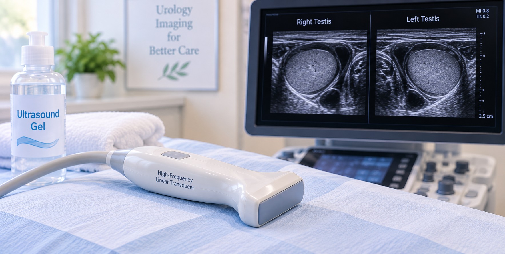
Understanding Your Testicular and Scrotal Ultrasound
A testicular and scrotal ultrasound provides important information about your testicles, epididymis, and surrounding scrotal tissues. It uses sound waves to create clear images, with no radiation or needles. At Sound Imaging, a scrotal ultrasound can help your healthcare provider quickly and accurately identify the cause of pain, swelling, or a lump.
A scrotal ultrasound shows things a physical exam can’t. Using Doppler imaging to assess blood flow, these images can detect testicular masses, infections, fluid buildup, enlarged veins, and testicular torsion. They help your provider understand your symptoms, evaluate fertility concerns, and make informed decisions about your care.


Depending on your symptoms and the reason for your exam, a testicular and scrotal ultrasound may be used to:
- Check for a lump or mass in the testicle or scrotum
- Find the cause of testicular pain or swelling
- Check blood flow to rule out testicular torsion
- Look for infection or inflammation, such as epididymitis or orchitis
- Find fluid buildup, cysts, or enlarged veins
- Locate an undescended testicle
- Provide more imaging when your healthcare provider recommends it
Different Testicular and Scrotal Ultrasounds
Scrotal Ultrasound
A complete scrotal ultrasound examines both testicles, the epididymis and the scrotal wall. It gives your provider a full view when you have pain, swelling or a change you can feel.
Scrotal Doppler Ultrasound
A scrotal Doppler ultrasound measures blood flow to the testicles. It is the main test for testicular torsion, a twisted testicle that cuts off its blood supply and needs emergency care. It also shows the increased blood flow seen with infection.
Testicular Mass Ultrasound
A testicular mass ultrasound looks closely at a lump found in or near the testicle. It can show whether the lump is inside the testicle, which may need further testing for testicular cancer, or outside it, where most lumps are harmless.
Epididymitis and Orchitis Ultrasound
This ultrasound checks for swelling and inflammation of the epididymis or testicle, often caused by infection. It helps explain pain, tenderness and swelling that develop over hours to days.
Hydrocele Ultrasound
A hydrocele ultrasound finds fluid collecting around the testicle, which causes painless scrotal swelling. It confirms the swelling is fluid and checks that the testicle underneath looks healthy.
Varicocele Ultrasound
A varicocele ultrasound finds enlarged veins in the scrotum, similar to varicose veins. Varicoceles can cause a dull ache or heaviness and are a common, treatable cause of male infertility.
Spermatocele and Epididymal Cyst Ultrasound
This ultrasound checks fluid-filled cysts in the epididymis, the tube behind the testicle. These cysts are usually harmless, and ultrasound helps tell them apart from other lumps.
Male Infertility Ultrasound
A male infertility ultrasound checks testicle size and texture and looks for varicoceles. Along with a semen analysis, it helps your provider or urologist find treatable causes of infertility.
Inguinal Hernia Ultrasound
An inguinal hernia ultrasound checks for bowel or fatty tissue pushing into the groin or scrotum. Your sonographer may ask you to cough or strain so the hernia can be seen.
Scrotal Trauma Ultrasound
A scrotal trauma ultrasound checks for injury after a blow to the groin. It can find a ruptured testicle, bleeding or blood collections that may need prompt treatment.
Undescended Testicle Ultrasound
An undescended testicle ultrasound looks for a testicle that has not moved into the scrotum, most often in babies and young boys. It can locate a testicle in the groin to help with treatment planning.

What to Expect
If You Have Sudden, Severe Testicular Pain
-
Sudden, severe pain in the testicle can be a sign of testicular torsion, a medical emergency. Do not wait for a scheduled appointment. Go to the nearest emergency room or call 911 right away.
Before your Appointment
-
You will be asked to undress from the waist down and will be given a gown or sheet to keep you covered. You are welcome to ask for a chaperone at any time.
- Your preparation may vary depending on the reason for your exam. When you schedule your appointment, our staff will let you know about any special instructions. For most testicular and scrotal ultrasounds, no special preparation is needed.
When You Arrive
- Our team will check you in and guide you to the ultrasound room. Your sonographer will explain the exam and help you get comfortably positioned before the study begins.
During Your Ultrasound
- A registered sonographer will bring you to a private exam room and have you lie on your back on the exam table. For privacy, only the area being examined is uncovered. A towel is placed under the scrotum for support, and the penis is covered with a towel or sheet.
- Your sonographer will apply warm gel to the scrotum and gently move a small handheld transducer over the area. Most patients feel only light pressure. If you have pain or tenderness, let your sonographer know, and they will be as gentle as possible. Doppler imaging is used to assess blood flow to your testicles and may produce a soft whooshing sound. This is normal.
- If your provider is checking for a varicocele or hernia, your sonographer may ask you to take a deep breath and bear down, as if having a bowel movement, or to stand for a few images. This helps enlarged veins or a hernia show more clearly.
How Long Will It Take?
- Most testicular and scrotal ultrasounds take 20 to 30 minutes. A varicocele or hernia evaluation may take a little longer because images are taken in more than one position.
After Your Exam
- There is no recovery time. The gel wipes off easily, and you can drive, go to work and return to your normal activities right away.
Getting Your Testicular Ultrasound Results
- A radiologist will review your scrotal ultrasound images and send a report to your healthcare provider, usually within a few business days. Your provider will contact you to explain your results and talk about next steps, such as antibiotics, follow-up imaging or a referral to a urologist. Your sonographer is not able to share results during the exam. If a finding needs quick attention, such as reduced blood flow or a concerning mass, your provider will be contacted right away.
FAQ
Does a testicular ultrasound hurt?
No. Most patients feel only light pressure from the transducer. If the area is swollen or tender, your sonographer will go slowly and be as gentle as possible.
Do I need to do anything to prepare for a scrotal ultrasound?
Usually not. You can eat, drink and take your medicine as normal, and you do not need a full bladder.
Can a testicular ultrasound detect cancer?
A testicular ultrasound can find masses in the testicle and show whether they are solid or fluid-filled. If a solid mass is found, your provider will order more testing to find out whether it is cancer.
Can I request a male sonographer or a chaperone?
Yes. Let our staff know when you schedule, and we will do our best to meet your request. You can ask for a chaperone at any time.
Is a scrotal ultrasound safe?
Yes. Ultrasound uses sound waves instead of radiation and is safe for patients of all ages, including children.
Insurance
Sound Imaging is in network with
Aetna
Blue Cross
Cigna
Meritain Health
Moda Health
TriWest
UnitedHealthcare
Alaska Medicaid including Denali KidCare
Medicare (coming soon)
Coverage and benefits vary by individual insurance plan. Questions about your coverage? Call (907) 531-2726.
No Surprise Act
Public Health Service (PHS) Act 2799B-3 and Section 45 of the Code of Federal Regulations (CFR) 149.430, describe medical consumer legal protections known as the No Surprise Act (NSA).
If you have an emergency medical condition and get emergency services from an out of network provider or facility, the most the provider or facility may bill you is your plan’s in network cost-sharing amount (such as copayments and coinsurance). You can’t be balance billed for these emergency services.
No Surprise Act Good Faith Estimates
Upon request, we are happy to provide registered patients with a good faith estimate based upon physician prescribed services, which will include expected charges for items or services as published in our current standard fee schedule. The estimate is based on information known at the time the estimate was created. The Good Faith Estimate does not include any unknown or unexpected costs that may arise during treatment. You could be charged more if complications or special circumstances occur.