
Vascular Ultrasound
Sound Imaging provides vascular and Doppler ultrasounds in Anchorage and Soldotna, Alaska, to evaluate blood flow through your arteries and veins. These exams help detect blood clots, including deep vein thrombosis (DVT), narrowed arteries, and aneurysms.
A carotid ultrasound evaluates narrowing in the neck arteries that may increase stroke risk. An arm or leg venous ultrasound checks for blood clots that may cause pain or swelling. If your doctor has recommended a vascular ultrasound, contact Sound Imaging to book your appointment.

Understanding Your Vascular Ultrasound
A vascular ultrasound, also called a Doppler ultrasound, shows how blood flows through the arteries and veins in your neck, arms, and legs. At Sound Imaging, a vascular ultrasound can help your healthcare provider find blood clots, blockages, and other circulation problems.
A carotid ultrasound can reveal early signs of stroke risk. A venous duplex scan can find deep vein thrombosis (DVT). An arterial Doppler test can check for peripheral artery disease (PAD) or aneurysms. These images help your provider explain leg pain, swelling, or numbness and plan your treatment.


Depending on the reason for your exam, a vascular ultrasound may be used to:
- Evaluate blood flow through arteries and veins
- Check for deep vein thrombosis (DVT) or other blood clots
- Detect narrowed or blocked arteries
- Evaluate circulation in the arms or legs
- Examine the carotid arteries in the neck
- Assess blood flow following vascular procedures
- Screen the abdominal aorta for an abdominal aortic aneurysm (AAA)
Types of Vascular Ultrasound Exams
Carotid Ultrasound
Carotid ultrasound uses Doppler imaging to evaluate blood flow in the carotid arteries in your neck that supply your brain. This noninvasive exam helps identify plaque buildup and artery narrowing that may increase stroke risk.
Arm-Venous Ultrasound
Arm venous ultrasound uses Doppler imaging to evaluate blood flow in the veins of your arm. Also called an upper-extremity venous ultrasound, this exam helps detect blood clots, including deep vein thrombosis (DVT), and to investigate arm pain or swelling.
Leg Venous Ultrasound
Leg venous ultrasound uses Doppler imaging to evaluate blood flow in your leg veins. Also called a lower-extremity venous ultrasound, this exam helps detect blood clots, including deep vein thrombosis (DVT), and to investigate leg pain or swelling.

What to Expect
Before your Appointment
-
Preparation for a vascular ultrasound depends on the type of exam being performed. Most vascular studies require little or no special preparation. For certain abdominal vascular ultrasound exams, you may be asked to avoid eating or drinking for several hours beforehand. You will receive specific instructions when your exam is scheduled.
- Please wear comfortable clothing and bring any information or paperwork requested by your provider.
When You Arrive
- Our team will check you in and take you to the ultrasound room. Your sonographer will explain the vascular ultrasound exam and help position you comfortably. You may be asked to change into a gown depending on the area being examined.
During Your Ultrasound
-
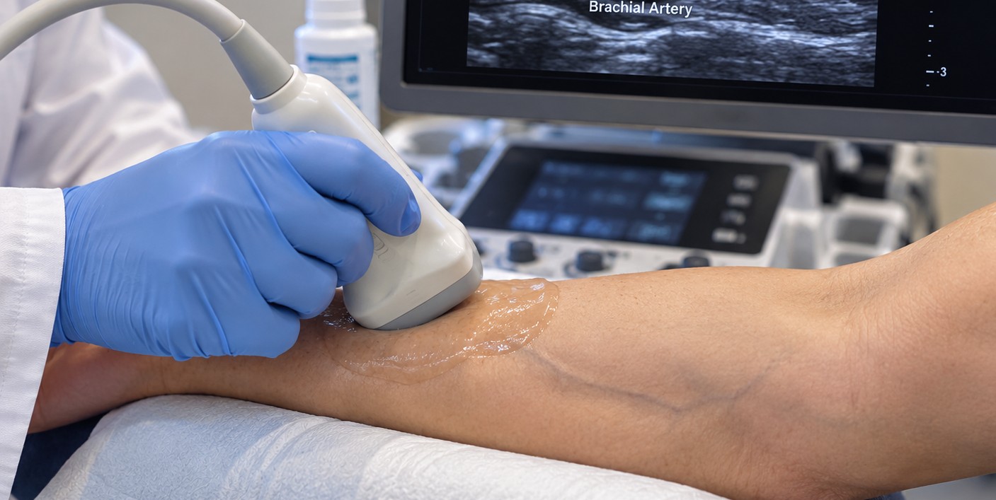
A small amount of ultrasound gel will be applied to the area being examined. The sonographer will gently move a handheld device called a transducer over your skin to create images of your arteries or veins.
-
Doppler ultrasound may also be used to evaluate the direction and speed of blood flow. You may hear pulsing or whooshing sounds from the ultrasound equipment as blood flow is measured. Some exams require gentle pressure with the transducer to evaluate the veins.
How Long Will It Take?
- Most vascular ultrasound exams take approximately 30–60 minutes. The length of your appointment may vary depending on the type of vascular study and the number of areas that need to be examined.
After Your Exam
-
Once the exam is complete, the gel will be removed. You can usually return to your normal activities immediately.
-
Your vascular ultrasound images and measurements will be reviewed, and the results will be sent to your referring healthcare provider. Your provider will discuss the findings and any recommended next steps with you.
Getting Your Vascular Ultrasound Results
- After your vascular ultrasound, a radiologist will review your images and blood flow measurements. They look for blood clots, narrowed or blocked arteries, plaque buildup, and problems with the valves in your veins. A report is then sent to your healthcare provider, usually within a few business days. Your provider will contact you to explain your Doppler ultrasound results and discuss next steps, such as medication, lifestyle changes, follow-up imaging, or a referral to a vascular specialist. If the ultrasound reveals a serious finding, such as deep vein thrombosis (DVT), we will contact your healthcare provider immediately so you can receive prompt care.
FAQ
What does a vascular ultrasound check for?
A vascular ultrasound evaluates blood flow through the arteries and veins. It can help detect blood clots, narrowed or blocked arteries, poor circulation, venous insufficiency and other vascular conditions. It may also be used to evaluate the carotid arteries or screen the abdominal aorta for an aneurysm.
Can an ultrasound detect a blood clot in the leg?
Yes. A venous ultrasound is commonly used to look for deep vein thrombosis (DVT), a blood clot that develops in a deep vein, usually in the leg. The sonographer may gently compress different areas of the vein while using ultrasound and Doppler imaging to evaluate blood flow.
How long does a vascular ultrasound take?
Most vascular ultrasound exams take approximately 30–60 minutes. The exact time depends on the type of vascular study, the area being examined and whether one or both sides of the body need to be evaluated.
Do I need to prepare for a vascular ultrasound?
Most vascular ultrasound exams require little or no preparation. For an abdominal vascular ultrasound, you may be instructed not to eat or drink for several hours before your appointment. Follow the preparation instructions provided for your specific exam.
Is a vascular ultrasound painful?
Insurance
Sound Imaging is in network with
Aetna
Blue Cross
Cigna
Meritain Health
Moda Health
TriWest
UnitedHealthcare
Alaska Medicaid including Denali KidCare
Medicare (coming soon)
Coverage and benefits vary by individual insurance plan. Questions about your coverage? Call (907) 531-2726.
No Surprise Act
Public Health Service (PHS) Act 2799B-3 and Section 45 of the Code of Federal Regulations (CFR) 149.430, describe medical consumer legal protections known as the No Surprise Act (NSA).
If you have an emergency medical condition and get emergency services from an out of network provider or facility, the most the provider or facility may bill you is your plan’s in network cost-sharing amount (such as copayments and coinsurance). You can’t be balance billed for these emergency services.
No Surprise Act Good Faith Estimates
Upon request, we are happy to provide registered patients with a good faith estimate based upon physician prescribed services, which will include expected charges for items or services as published in our current standard fee schedule. The estimate is based on information known at the time the estimate was created. The Good Faith Estimate does not include any unknown or unexpected costs that may arise during treatment. You could be charged more if complications or special circumstances occur.