
High-Risk Pregnancy & MFM
Sound Imaging offers high-risk pregnancy ultrasound in Anchorage and Soldotna, Alaska, with exams interpreted by Maternal-Fetal-Medicine (MFM) specialists, also called perinatologists. We assess your baby’s growth, development, and well-being, providing your provider with information to guide your care.
Close monitoring may be recommended for multiples, diabetes, high blood pressure, maternal age of 35 or older, previous pregnancy with complications, or concerns on an earlier ultrasound. Depending on your provider’s order, exams may include a detailed fetal anatomy scan, fetal echo, Doppler blood flow studies, cervical lenght measurements, and biophysical profiles (BPP’s).

Understanding Your High-Risk Pregnancy Ultrasound
When you or your baby needs closer monitoring, Sound Imaging provides high-risk pregnancy ultrasound with personalized care. Detailed images of your baby, placenta, and amniotic fluid are reviewed by a Maternal-Fetal-Medicine (MFM) specialist, helping your healthcare team understand your baby’s development, evlauate concerns, and guide your care throughout your pregnancy.
A high-risk pregnancy ultrasound provides a detailed look at your baby’s growth and well-being. Depending on your needs, the exam may evaluate your baby’s anatomy, heart, blood flow, and movements, or measure your cervical length. The results help your provider monitor your pregnancy and guide the next steps in your care.


Depending on your health history and the reason for your exam, a high-risk pregnancy ultrasound may be used to:
- Take a detailed look at your baby’s anatomy and organs
- Check your baby’s heart structure, rhythm, and function.
- Track your baby’s growth
- Evaluate blood flow in the umbilical cord and your baby’s brain
- Monitor twins, triplets, and other multiple pregnancies
- Check the placenta, amniotic fluid, and cervical length
- Check your baby’s well-being with a biophysical profile
- Provide more imaging when your provider or MFM specialist recommends it
Different High-Risk Pregnancy Ultrasounds

Targeted Anatomy Scan (Level II Ultrasound)
A targeted anatomy scan, also called a Level II ultrasound, is a more detailed version of the standard 20-week scan. It takes a close look at your baby’s brain, heart, spine, face, kidneys and limbs when there is a higher risk of birth defects or a concern from an earlier ultrasound.
Fetal Echo
A fetal echocardiogram is a specialized ultrasound of your baby’s heart. In a high-risk pregnancy, it is often recommended for diabetes, IVF pregnancies, a family history of heart defects or an abnormal finding on the anatomy scan, so any heart condition can be planned for before birth.
Serial Growth Ultrasounds
Serial growth ultrasounds measure your baby’s size every few weeks. They help find fetal growth restriction (FGR) or a larger-than-expected baby, and are often recommended for diabetes, high blood pressure, preeclampsia or a previous small baby.
Umbilical Artery Doppler
An umbilical artery Doppler measures blood flow through the umbilical cord. It shows how well the placenta is delivering oxygen and nutrients, and helps your MFM specialist decide how often to monitor a baby with growth restriction.
Middle Cerebral Artery (MCA) Doppler
An MCA Doppler measures blood flow in an artery in your baby’s brain. It is used to check for fetal anemia, especially when there are red blood cell antibodies, and to monitor babies with growth problems.
Biophysical Profile (BPP)
A biophysical profile checks your baby’s breathing movements, body movement, muscle tone and amniotic fluid. In a high-risk pregnancy, it is often done weekly or twice weekly in the third trimester to confirm your baby is doing well.
Cervical Length Monitoring
Cervical length monitoring uses repeat transvaginal ultrasounds to measure your cervix. It is recommended for patients with a history of preterm birth, cervical surgery or a short cervix, so treatment such as progesterone or a cerclage can be considered early.
Twin and Multiple Pregnancy Ultrasound
Twin and multiple pregnancy ultrasounds check each baby’s growth, amniotic fluid and position. For twins who share a placenta, frequent scans watch for twin-to-twin transfusion syndrome (TTTS) and other complications that need early care.
Placenta Evaluation Ultrasound
A placenta evaluation ultrasound checks the location and health of your placenta. It can find placenta previa, where the placenta covers the cervix, and signs of placenta accreta, which is more common after a previous C-section.
Amniotic Fluid Assessment
An amniotic fluid assessment measures the fluid around your baby. Too little fluid (oligohydramnios) or too much fluid (polyhydramnios) can be linked to diabetes, growth problems or other conditions that need closer monitoring.
Nuchal Translucency and Early Risk Assessment
A nuchal translucency scan between 11 and 14 weeks measures fluid at the back of your baby’s neck. In a high-risk pregnancy, it gives early information about chromosomal conditions and heart defects, helping your MFM specialist plan further testing.

What to Expect
Before your Appointment
-
Bring your photo ID, your insurance card and your OB-GYN or MFM specialist’s order. Your preparation may vary depending on the type of ultrasound you are having. When you schedule your appointment, our staff will let you know if there are any special instructions, such as drinking water to arrive with a full bladder or avoiding food for a certain period of time.
- Wear comfortable, two-piece clothing so your belly can be easily uncovered. You are welcome to bring a support person. Ask our staff how many guests are allowed in the exam room.
When You Arrive
- Our team will check you in and guide you to the ultrasound room. Your sonographer will explain the exam and help you get comfortably positioned before the study begins.
During Your Ultrasound
-
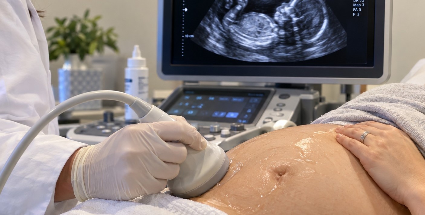
A registered sonographer with experience in high-risk and MFM imaging will bring you to a private exam room. You will lie on the exam table, and your sonographer will apply warm gel on your abdomen and gently move a small handheld transducer across your skin.
Your sonographer will take careful measurements of your baby’s growth, anatomy, heart, placenta, amniotic fluid, and document fetal movement. Doppler imaging may be used to check blood flow in the umbilical cord or your baby’s brain. This can make a soft whooshing sound, which is normal. If your baby is not in a good position, you may be asked to roll onto your side, walk for a few minutes, or empty your bladder.
Some exams, such as cervical length monitoring or an early pregnancy scan, may include a transvaginal ultrasound for clearer images. Most patients feel only mild pressure. You can ask for a chaperone at any time.
How Long Will It Take?
- Most high-risk pregnancy ultrasounds take 60 minutes. A targeted anatomy scan (Level II ultrasound), a fetal echocardiogram, or a twin pregnancy ultrasound may take 60 to 90 minutes. A biophysical profile usually takes about 30 minutes.
After Your Exam
- There is no recovery time. You can eat, drive and return to your normal activities right away. Because high-risk pregnancies often need regular monitoring, our staff can help you schedule your next ultrasound before you leave.
We’re Here to Make Your Visit Comfortable
- A maternal-fetal medicine specialist reviews your ultrasound images and sends a report to your provider, who will explain the results and discuss next steps with you. These may include more frequent monitoring, additional testing, genetic counseling, or delivery planning. Your sonographer can show you your baby on the screen and explain the imaging process but cannot provide medical results during the exam. If a finding needs urgent attention, we will contact your provider right away.
FAQ
Why do I need more ultrasounds in a high-risk pregnancy?
Conditions like diabetes, high blood pressure, preeclampsia, or carrying twins can affect your baby’s growth and well-being. More frequent ultrasounds help your care team catch changes early and keep you and your baby safe.
What is the difference between a regular ultrasound and a Level II ultrasound?
A Level II ultrasound, also called a targeted anatomy scan, is a more detailed exam. It takes longer and looks more closely at your baby’s organs, heart and development than a standard anatomy scan.
How often will I need a high-risk pregnancy ultrasound?
It depends on your condition. Some patients need an ultrasound every 3 to 4 weeks for growth, while others need weekly or twice-weekly exams.
Is it safe to have many ultrasounds during pregnancy?
Yes. Ultrasound uses sound waves instead of radiation and has been used safely in pregnancy for decades.
Can I find out fetal sex during a high-risk pregnancy ultrasound?
Often, yes. Let your sonographer know if you want to know, or if you would like to keep it a surprise.
Insurance
Sound Imaging is in network with
Aetna
Blue Cross
Cigna
Meritain Health
Moda Health
TriWest
UnitedHealthcare
Alaska Medicaid including Denali KidCare
Medicare (coming soon)
Coverage and benefits vary by individual insurance plan. Questions about your coverage? Call (907) 531-2726.
No Surprise Act
Public Health Service (PHS) Act 2799B-3 and Section 45 of the Code of Federal Regulations (CFR) 149.430, describe medical consumer legal protections known as the No Surprise Act (NSA).
If you have an emergency medical condition and get emergency services from an out of network provider or facility, the most the provider or facility may bill you is your plan’s in network cost-sharing amount (such as copayments and coinsurance). You can’t be balance billed for these emergency services.
No Surprise Act Good Faith Estimates
Upon request, we are happy to provide registered patients with a good faith estimate based upon physician prescribed services, which will include expected charges for items or services as published in our current standard fee schedule. The estimate is based on information known at the time the estimate was created. The Good Faith Estimate does not include any unknown or unexpected costs that may arise during treatment. You could be charged more if complications or special circumstances occur.